
The 2D-to-3D Registration Bottleneck in Computer-Assisted Surgery
Recovering an accurate spatial transform between a patient's preoperative 3D anatomy and live surgical tools has been the fundamental stumbling block of image-guided interventions for decades. Mathematically, the problem is an unconstrained 2D-to-3D rigid registration challenge: given a calibrated 2D X-ray projection $I_{2D} \in \mathbb{R}^{H \times W}$ acquired intraoperatively by a fluoroscopic C-arm, you must recover the 6-degree-of-freedom (6-DoF) SE(3) transformation matrix $[R \mid t]$ that aligns the coordinate system of the preoperative volumetric CT or MR scan $V_{3D} \in \mathbb{R}^{D \times H \times W}$ to the projector’s physical frame.
In an active operating room, this translation from 2D shadow to 3D orientation forces an operational compromise that I have watched teams struggle with repeatedly. Classical image-guided surgery divides into two equally flawed camps:
First, pure image-based iterative registration. Systems in this category project synthetic X-rays—known as Digitally Reconstructed Radiographs (DRRs)—through the 3D CT volume via ray-casting, compare the synthetic projection to the physical fluoroscopic image using handcrafted similarity metrics like Normalized Cross-Correlation or Mutual Information, and execute an iterative search over parameter space. Because standard ray-casting through dense voxel grids is computationally punishing, computing gradients or sampling across rotational and translational axes takes tens of seconds to multiple minutes per frame. In orthopedic or spinal interventions where surgeons adjust hardware continuously, a sixty-second latency loop destroys surgical flow and invalidates the real-time premise.
Second, the optical tracker workaround. To bypass ray-casting latency, commercial navigation systems like Medtronic StealthStation or Brainlab platforms rely heavily on physical optical tracking arrays. Rigid tracking frames studded with retroreflective spheres are drilled directly into the patient's bone (such as the iliac crest or spinal process) and registered via paired-point matching. This shifts the tracking burden from image computation to external infrared cameras. Yet optical setups introduce their own failure modes: line-of-sight occlusions by operating staff, mechanical deflection of tracker pins, cumbersome sterile drape management, and invasive secondary incision sites.
What clinical engineering has demanded is markerless, instantaneous registration: bringing a fluoroscopy C-arm into position, depressing the pedal, and immediately seeing the volumetric CT correctly overlaid within sub-millimeter Target Registration Error (TRE). Closing that gap without relying on fragile external hardware has been the holy grail of spatial surgical navigation—a challenge analogous to the persistent coordinate anchoring problems we see in generalized spatial pipelines, which I analyzed when examining long-horizon dense 3D tracking architectures.
The Two-Phase Architecture: 5-Minute Pre-Op Synthesis Meets Sub-Second Pose Estimation
The core architectural innovation of xvr—developed by Vivek Gopalakrishnan and collaborators—is recognizing that 2D-to-3D registration should not be treated as a cold-start intraoperative search, nor as an over-generalized population-scale regression problem. Instead, xvr decouples the registration pipeline into two structurally distinct phases: an offline patient-specific adaptation phase, followed by an ultra-fast intraoperative inference loop.
sequenceDiagram
    autonumber
    participant PACS as Hospital PACS
    participant EdgeGPU as On-Prem GPU Worker
    participant DiffDRR as Differentiable Ray-Caster
    participant CArm as Intraoperative C-Arm
    participant XVR as xvr Inference Engine
    participant Nav as Surgical Navigation Display

    Note over PACS,EdgeGPU: Phase 1: Preoperative Patient Adaptation (~5 Mins)
    PACS->>EdgeGPU: Ingest Pre-op Volumetric CT / MR (DICOM)
    loop Self-Supervised Geometry Adaptation
        EdgeGPU->>DiffDRR: Sample SE(3) Pose Perturbations
        DiffDRR-->>EdgeGPU: Synthesize Differentiable DRRs
        EdgeGPU->>EdgeGPU: Update Patient-Specific Foundation Weights
    end
    EdgeGPU->>XVR: Deploy Calibrated Patient Checkpoint

    Note over CArm,Nav: Phase 2: Intraoperative Low-Latency Tracking (Sub-Second)
    CArm->>XVR: Stream Live 2D Fluoroscopy Projection (DICOM-RTI)
    XVR->>XVR: Forward Pass Pose Regression via Calibrated Backbone
    XVR->>Nav: Stream 6-DoF Rigid Matrix [R | t]
    Nav-->>Nav: Overlay Volumetric CT on Live Fluoroscopy Stream
The historical failure of generalized deep networks in medical registration stemmed from the sheer diversity of human anatomy, surgical positioning, and tissue density variations. A network trained to regress pose on five hundred patient pelvises inevitably stumbles when presented with a dysplastic femur, severe osteophytes, or abnormal bone mineral density. xvr sidesteps this generalization trap through forward simulation.
During the preoperative phase—taking roughly five minutes on local hardware while the patient is prepped and draped—the system ingests the patient’s volumetric CT from PACS. Rather than relying on static anatomical assumptions, xvr uses hardware-accelerated, auto-differentiable DRR projection (leveraging the DiffDRR framework) to simulate thousands of prospective projective geometries directly from that specific scan. By sampling camera intrinsic geometries, focal lengths, and rotational bounds, it creates a self-supervised training manifold tailored exclusively to that individual's density distribution.
When the patient is on the operating table and the C-arm fires, the compute requirements invert. The intraoperative module does not need to compute hundreds of ray-cast passes or resolve high-dimensional optimization landscapes under active sterile field pressure. The adapted neural backbone processes the incoming 2D projection and directly estimates the 6-DoF matrix in fractions of a second. This hybrid approach turns differentiable physics into an offline training supervisor rather than an intraoperative bottleneck, preserving sub-millimeter anatomical precision while delivering sub-second frame rates directly to the surgical monitor.
Evaluating the Competitive Landscape: Classical Ray-Casting vs Deep Regressors vs xvr
Navigating 2D-to-3D registration requires balancing two competing metrics: intraoperative execution speed and anatomical generalization robustness. When mapping where modern clinical approaches sit on this frontier, the historical trade-offs become glaringly apparent.
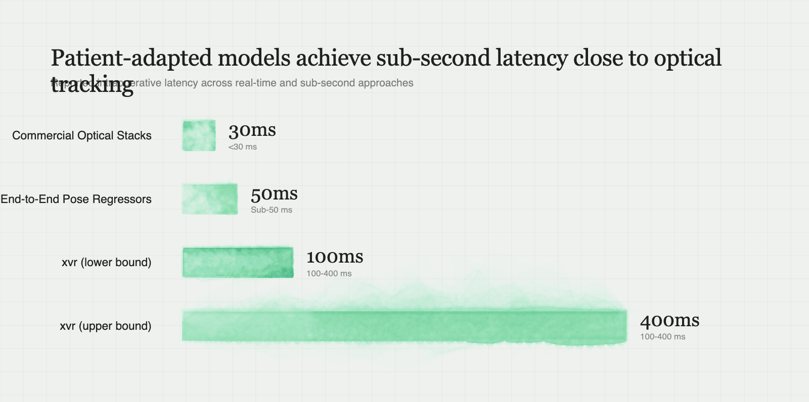
Classical software stacks such as the Insight Toolkit (ITK) and Plastimatch optimize DRR projections iteratively against target radiographs. Their mathematical foundations are unassailable and their deterministic properties make them attractive for regulatory clearance. However, their position in the clinical workflow is severely constrained: ray-marching through a $512 \times 512 \times 512$ voxel cube across hundreds of gradient descent steps takes between 15 to 90 seconds per fluoroscopic shot. That speed profile relegates iterative DRR alignment to static confirmation checkpoints rather than dynamic surgical navigation.
In response, computer vision researchers attempted end-to-end regression using convolutional pose backbones. Models in the PoseNet lineage regress the six Euler angles and translation vectors directly from 2D pixel intensities. While these architectures deliver millisecond-level forward passes, their clinical robustness is notoriously poor. Direct regressors learn spurious correlation shortcuts based on image borders, patient soft-tissue contours, or table radiopacity. The moment a real-world surgical artifact appears—such as a radiopaque surgical clamp, a radiolucent carbon-fiber table edge, or an unexpected variation in beam energy—direct pose regressors lose alignment entirely, exhibiting multi-centimeter tracking failures with no internal mechanism to gauge uncertainty.
Pure differentiable rendering engines like DiffDRR shifted the paradigm by exposing exact analytical gradients through the rendering pipeline. This represented a substantial leap over classical finite-difference ray-casting, compressing optimization runs down to single-digit seconds. Yet pure optimization still leaves the surgeon waiting several seconds per verification, which remains too slow for continuous, real-time procedural guidance.
| Architecture / Platform | Intraoperative Latency | Patient Generalization | Pre-Op Compute Overhead | Sensitivity to Surgical Occlusions | Tracking Hardware Dependency |
|---|---|---|---|---|---|
| Classical Iterative DRR (ITK, Plastimatch) | 15–90 seconds per shot | High (optimizes per scan without priors) | Negligible (volume loaded into memory) | Moderate (can trap in local minima from occluders) | Markerless (Purely radiograph-based) |
| Commercial Optical Stacks (Medtronic Stealth, Brainlab) | Real-time (<30 ms) | Complete (independent of internal anatomy) | Negligible (paired-point fiducial registration) | High (physical line-of-sight blockage of IR trackers) | Rigid bone pins, optical tracking arrays, IR cameras |
| End-to-End Pose Regressors (Direct CNN / PoseNet) | Sub-50 ms | Very Poor (fails on unseen anatomies & foreign bodies) | None (pre-trained population model) | Catastrophic (implants break feature maps) | Markerless |
| Pure Differentiable DRR (Standalone DiffDRR) | 2–8 seconds | High (exact analytical ray-casting) | Negligible (builds projector graph) | Moderate (susceptible to non-convex loss valleys) | Markerless |
Patient-Adapted Foundation (xvr) |
Sub-second (100–400 ms) | High (calibrated to patient volume in pre-op) | ~5 minutes local GPU forward simulation | Low (physics-based manifold regularizes features) | Markerless |
By dedicating five minutes of background compute to simulate the forward physics of the patient’s scan prior to the first incision, xvr bridges the gap. It captures the sub-millimeter stability of physics-based projection matching while unlocking the inference latency necessary for live intraoperative guidance.
The Limits of Rigid 6-DoF: Anatomical Shift, Spinal Flexion, and Soft Tissue Realities
The foundational premise of rigid registration is that the anatomical target moves through space as an unyielding solid. For monolithic bony structures—an intact cranium, an isolated femoral shaft, or an osteotomized pelvis held in a rigid clamp—that premise holds. In production surgical navigation, however, human anatomy rarely behaves like a machined aerospace component, and treating the patient as a single 6-DoF rigid transform introduces severe spatial error the moment you move past isolated long bones.
The most pervasive failure mode I watch teams encounter is posture discrepancy between preoperative acquisition and the surgical theater. A diagnostic CT is almost universally captured with the patient supine on a flat scanner couch. Orthopedic and neurosurgical spine interventions, by contrast, frequently place the patient prone on padded bolsters or an articulated Jackson table. That positional inversion immediately alters the physical curvature of the spine. Each vertebral level functions as a distinct rigid body connected by flexible intervertebral discs and facet joints. Under prone positioning, lumbar lordosis and thoracic kyphosis shift by several degrees per segment.
If you attempt to register an L1–L5 spinal segment using a global 6-DoF rigid assumption, the optimization landscape compromises across the entire chain. The model finds a mathematical average pose where no single vertebra is accurately aligned. A rotational mismatch of three degrees across an articulated segment translates into multiple millimeters of displacement at the vertebral pedicle—an unacceptable margin when guiding a drill tap adjacent to the spinal cord. Similar breakdown occurs in orthopedic trauma, where intraoperative reduction maneuvers displace fractured bone fragments relative to their pre-injury CT positions, and in visceral surgery, where diaphragmatic respiration continuously deforms soft tissue structures like the liver and kidneys.
These edge boundaries define where rigid systems like base xvr must yield to kinematic and deformable frameworks. Articulated registration frameworks—such as PolyPose, which parameterizes multi-body kinematics across connected joints—model anatomical relationships as kinematic trees rather than monolithic transforms. When planning clinical deployment, drawing this architectural line early is essential: rigid 6-DoF foundation models deliver unmatched speed for fixed skeletal anatomy, but articulated or deformable graph formulations remain mandatory whenever intersegmental posture or soft-tissue compliance breaks the rigid body assumption.
Edge Realities in the Operating Room: Metal Artifacts, Gantry Sag, and Low-Dose Noise
Moving from synthetic bench benchmarks to live operating rooms exposes registration pipelines to physical environment degradations that clean forward-simulation models rarely anticipate. The surgical field is cluttered with high-density foreign bodies, mechanical hardware flex, and extreme photon starvation—all of which systematically distort projection geometry.
The primary algorithmic trap is the false local minimum induced by radiopaque instrumentation. An intraoperative fluoroscopic image rarely contains just virgin bone; it is crowded with stainless steel Hohmann retractors, titanium pedicle screws, K-wires, suction tips, and radiopaque surgical sponges. Because metallic hardware attenuates X-rays far more aggressively than cortical bone, it casts pitch-black projection shadows with steep gradient edges. Standard similarity metrics—and naive learned feature extractors—gravitate toward these high-contrast borders. In practice, the registration engine will lock onto the contour of a retractor blade and attempt to align it with an anatomical ridge in the pre-op CT, pulling the estimated pose centimeters off target.
Compounding this image-space corruption is physical mechanical deflection, commonly known as gantry sag. A surgical C-arm suspends an X-ray source and flat-panel detector on a cantilevered metal arc. As the C-arm rotates from an anteroposterior projection to steep lateral or oblique views, gravity exerts variable torque on the assembly. The physical distance between source and detector, along with the piercing point on the detector panel, deflects by several millimeters and fractional degrees relative to nominal factory calibration. When a differentiable projector assumes an idealized pinhole model, that physical sag produces a systematic geometric bias across the reconstructed volume.
Finally, intraoperative imaging operates under strict ALARA (As Low As Reasonably Achievable) radiation constraints. Continuous fluoroscopy drops beam current drastically, bathing the detector in severe quantum mottle and scattered radiation. This high-frequency Poisson noise obliterates fine trabecular bone patterns, leaving only faint cortical silhouettes visible through the grain.
Deploying resilient 2D-to-3D pipelines demands defensive engineering against these realities. Instrument-induced divergence must be countered with automated feature-based loss masking, segmenting high-attenuation foreign contours prior to similarity evaluation so foreign metal is excluded from the alignment loss. Mechanical sag requires dynamic calibration lookups tied to the physical rotation angles of the arm, while gantry encoder integration should seed the model’s initial pose hypothesis directly from hardware metadata, ensuring the inference engine searches within an anchored local basin rather than gambling on unconstrained space.
Building Production Medical Vision Systems: Safety Fallbacks and Edge Infrastructure
Deploying a patient-adapted model like xvr inside an active surgical theater forces an uncompromising reckoning with hospital network topology and patient safety boundaries. Academic proofs of concept frequently assume seamless access to elastic cloud clusters, but in clinical reality, streaming volumetric patient scans outside the hospital intranet is an immediate regulatory dead end. Under HIPAA and equivalent data privacy regimes, transmitting uncompressed multi-gigabyte thin-slice CT studies containing Protected Health Information across WAN boundaries introduces severe compliance liability alongside unpredictable network latency. If a PACS transfer stalls over an external connection, the five-minute pre-op adaptation window expands uncontrollably, delaying the entire surgical schedule.
The only viable production footprint is an on-premises edge computing appliance isolated within the hospital's local surgical VLAN. In my experience sizing these deployments, housing a dedicated edge workstation equipped with dual high-VRAM GPUs (such as 24GB+ RTX 4090s or RTX 6000 Ada cards) directly in or adjacent to the operating room provides the exact compute density required. This machine runs an embedded DICOM C-STORE and C-MOVE listener daemon that autonomously intercepts volumetric series scheduled for the day's theater list. The daemon ingests the DICOM files, strips non-essential metadata, maps coordinates from DICOM Patient (LPS) space to the virtual ray-caster’s camera coordinate frame, and executes the self-supervised adaptation loop entirely on local PCIe lanes well before the initial incision.
Yet hardware isolation only resolves the ingestion problem; the harder operational challenge is patient safety verification. In computer-assisted surgery, displaying a silently corrupted registration is catastrophic—guiding a pedicle screw or drill tap based on a hallucinated pose can cause irreversible neurological or vascular trauma. A production medical vision pipeline cannot operate on blind trust; it requires an explicit out-of-distribution (OOD) verification metric and a fail-safe fallback mechanism.
To validate registration integrity in sub-second time, the runtime must execute an immediate closed-loop verification pass after each forward inference. The system feeds the predicted 6-DoF pose back into the differentiable projector to synthesize an instantaneous verification DRR. It then evaluates a masked gradient correlation and multi-scale normalized cross-correlation against the live fluoroscopy frame, focusing strictly on high-gradient anatomical edges while masking out known instrument geometries. If the resulting alignment metric falls below an empirically calibrated confidence floor—or if residual gradient vectors reveal high spatial variance indicative of localized false minima—the navigation engine must immediately execute an automated safety lockout. Rather than rendering an unverified overlay, the system suppresses the 3D visualization, alerts the surgical team to the anatomical mismatch, and falls back to displaying unaugmented fluoroscopy until the operator either recalibrates the C-arm projection angle or provides a rough manual pose re-initialization.
Applied Implementations: Translating Differentiable Registration into Practical Tools
Translating the principles of differentiable projection and patient-adapted foundation models into production software opens up high-leverage architectural patterns for applied engineers building in surgical guidance and spatial robotics. The core insight—spending brief offline cycles to generate physics-grounded forward simulations so an edge network can perform instant real-time alignment—generalizes far beyond isolated research scripts.
For engineers designing intraoperative tracking pipelines, a prime reference architecture is a Real-Time Markerless Orthopedic Screw Trajectory Verifier. This system operates as a low-latency surgical guidance daemon that ingests intraoperative lateral and anteroposterior fluoroscopy frames, registers them against preoperative pelvic or spinal CTs via xvr, and continuously renders the planned drill trajectory directly over the live video stream. Mechanically, the pipeline links a DiffDRR PyTorch projection backend and patient-adapted weights to a low-latency C-arm frame grabber tapping SDI or NDI streams, pushing updated SE(3) pose coordinates to an OpenGL or Vulkan surgical display overlay while a background DICOM Query/Retrieve listener manages case data. The chief implementation trap here lies in instrument insertion: as dense titanium pedicle screws, K-wires, and steel retractors enter the field of view, raw image-similarity metrics collapse into severe false minima. Production implementations must incorporate mask-based loss weighting or robust high-level feature losses that segment and discount non-biological radiopaque geometry before computing alignment loss.
A tracking daemon is only as dependable as its spatial initialization, which makes an Autonomous C-Arm Gantry Calibration and Pose Initialization Service an essential companion module. Unconstrained 6-DoF global search over wide projective angles invites convergence failure. Engineers can resolve this by building a lightweight microservice that consumes C-arm gantry encoder telemetry and spatial metadata over a serial or CAN-bus hardware bridge, paired with a Modality Performed Procedure Step (MPPS) parser. This service computes rigid pose priors to seed the xvr initialization dispatch handler, narrowing the registration search space to a localized cone. Crucially, this microservice must maintain pre-computed mechanical sag lookup tables. Because physical C-arm gantries flex under gravity as they rotate across orbital and lateral angles, nominal source-to-detector distances and principal piercing points drift by several millimeters. Injecting real-time geometric correction matrices calculated from empirical sag curves prevents systematic coordinate offsets from corrupting the virtual camera model.
Underpinning these live intraoperative tools is the edge data lifecycle, which can be cleanly formalized as an On-Premises Preoperative Adaptation Worker for Hospital Surgical Suites. This architecture functions as an isolated edge orchestration pipeline designed to bridge hospital PACS and compute infrastructure without disrupting clinical routines. Operating within an isolated local network, the worker connects an open-source PACS interface like DCM4CHEE to an automated batch volume resampler, feeding a PyTorch CUDA container that executes the self-supervised xvr simulation loop on a dual-RTX local workstation. Once adapted, the patient-specific network weights are encrypted and staged in a local cache indexed to the surgical schedule identifier. The critical bottleneck to engineer around is the PACS retrieval boundary: pulling multi-gigabyte uncompressed thin-slice DICOM series synchronously at the start of a procedure triggers network congestion and data governance friction, easily blowing past the five-minute adaptation window. Structuring the worker to asynchronously pre-fetch and resample scheduled scans during pre-op prep ensures that calibrated weights are already warm the moment the patient is wheeled into the theater.
Resources
(no official sources were available to link)
Updated 2026-09-22 by Mehran Mozaffari.
Related posts
29 October 2026
How 3D OS Turns a Coding Agent into a Six-Department Animation Studio Without a GUI
15 September 2026
From Static Mesh to Walking Character: A Technical Operator's Manual for the 3D Vibe Coding Pipeline
23 June 2026
The Splat Stack: Where Gaussian Splatting Turns Into Five Different Products
8 June 2026
CADGenBench Grades AI-Generated Mechanical Parts Like an Engineer Would: A Validity Gate, Keep-In Volumes, and Betti Numbers
3 June 2026
From Pixels to Programs: What VIGA, Articraft, and OmniLottie Prove About Code-Native Visual AI
1 November 2026
In-Session Context Compaction or Isolated Subagent Threads for Long-Running Coding Agents